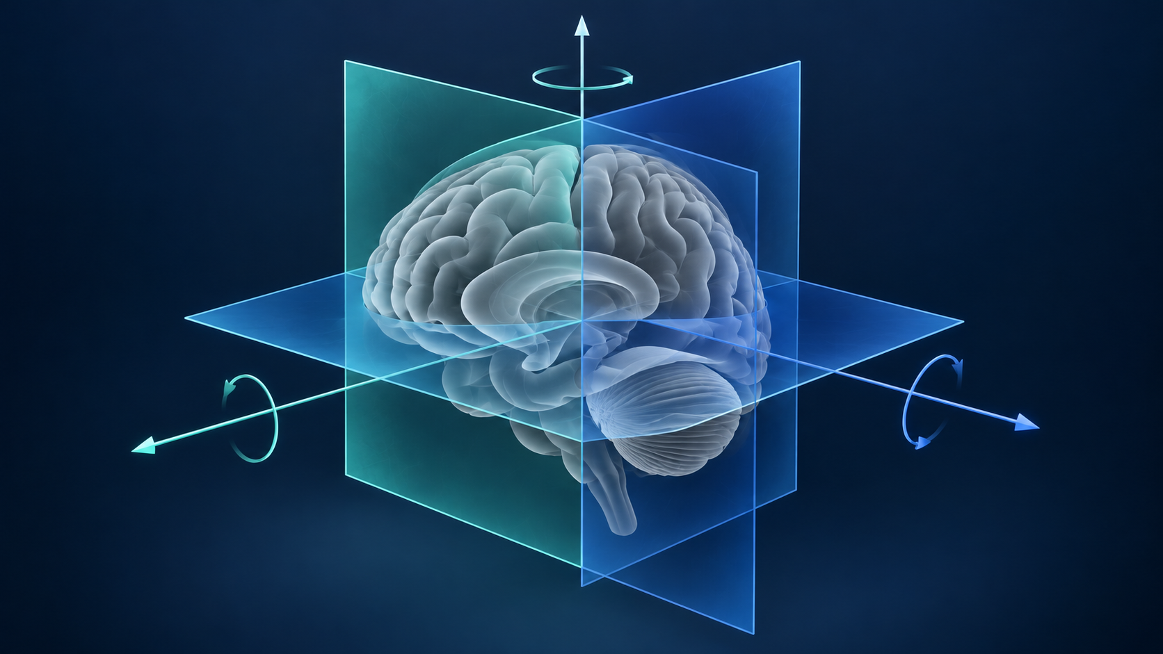
MIT researchers and collaborators at Boston Children’s Hospital and Inria have developed E(3)-Pose, an AI model that estimates fetal head orientation in MRI. MIT’s October 19 report describes a design that incorporates geometric symmetry into the model rather than expecting it to learn every possible orientation independently.
The work, presented at ECCV 2026, addresses motion that complicates the acquisition of aligned slices for three-dimensional reconstruction. Training focused primarily on third-trimester imaging; extending the approach to earlier gestation remains a research goal. The announcement does not establish improved patient outcomes or diagnostic reliability across all stages of pregnancy.
NAOAT analysis
An orientation estimate is an intermediate product. Its clinical usefulness depends on whether an imaging workflow can turn that estimate into adequate anatomical coverage before movement changes the situation again. A technically accurate prediction that arrives too late could still be difficult to use during acquisition.
That suggests a practical distinction between retrospective evaluation and acquisition testing. Replaying a recorded scan can assess whether the model recognizes position. A prospective workflow also has to determine how the scanner responds, whether the next slice covers the intended anatomy, and whether repeated corrections create gaps elsewhere in the volume.
Earlier gestation introduces another evaluation question: does the model retain useful behavior when anatomy differs from the examples on which it was developed? Treating that extension as a separate validation task would make its limits easier to understand than presenting fetal MRI as a single uniform setting.
The architectural idea is consequential because it gives the model an explicit rule about geometry. The next evidence to look for is a measured connection between that rule, acquisition performance, and usable reconstructed images. Those are distinct steps toward a clinical application, each deserving its own assessment.